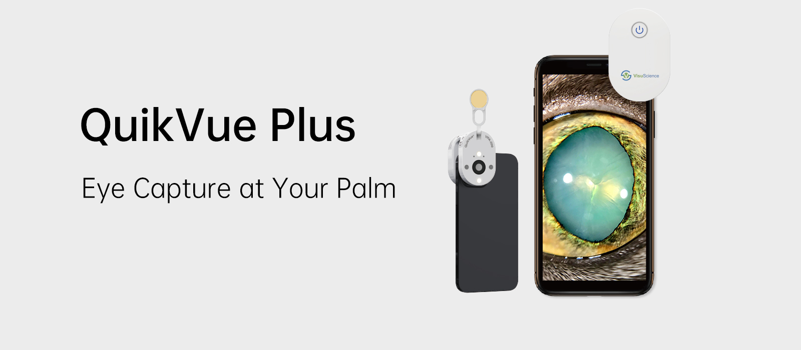

| QuikVue Plus Design Concept It is well acknowledged that imaging is playing a more and more important role in veterinary ophthalmology diagnostics. | |||||||
 | |||||||
| White and Blue Illumination | |||||||
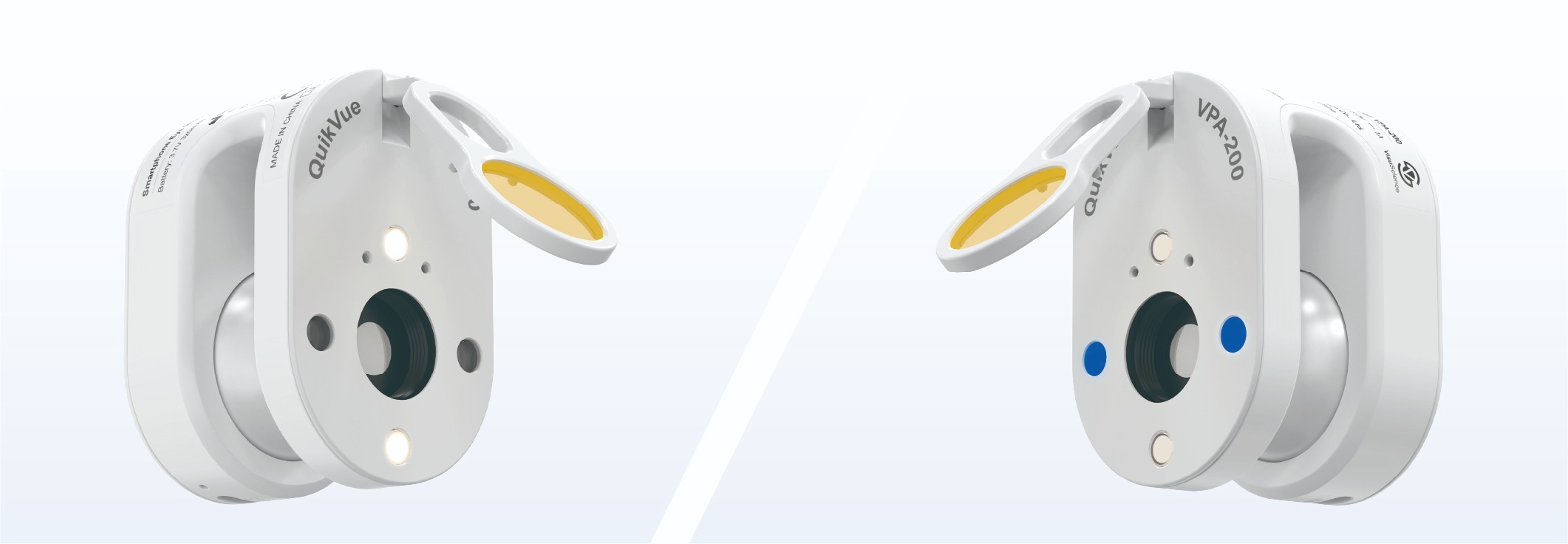
| QuikVue Plus provides both white and blue illumination respectively. The white LED projects warm white light which is similar to slit lamp’s halogen illumination. There are two levels of white illumination available to meet different brightness demand during examination. The blue illumination can be used to capture fluorescein images to assist diagnosis with corneal staining and contact lens fitting, etc. |  | ||||||
| Integrated Yellow Filter | |||||||
| QuikVue Plus is equipped with an integrated yellow filter. The flipable yellow filter making it easy for clinicians to take enhanced fluorescein images with cobalt blue illumination. This function is especially useful in lens fitting, cornea ulcer imaging, tear film break up time examination, etc. | ||||||
Smartphone Compatibility | |||||||
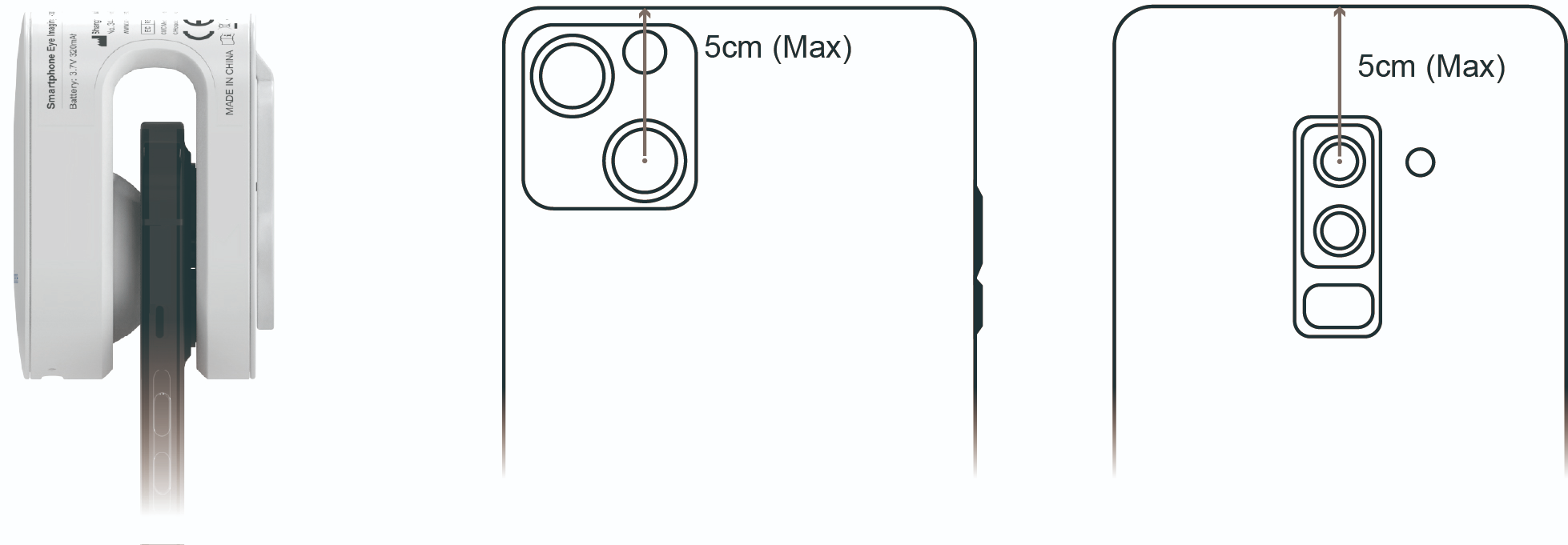
Thanks to the air cushion design, QuikVue Plus is able to attach on most of the phones in the market firmly. Clinicians need to align QuikVue Plus lens with smartphone’s main camera. The requirements for the main camera location is as below: | |||||||
 | |||||||
Working Application | |||||||
QuikVue plus can work with smartphone default camera app or VisuScience developed VisuDoc ophthalmic case management app. VisuDoc can be downloaded from both App store and Google play store. | |||||||
 | |||||||
| Power Management | |||||||
QuikVue Plus applies mini Li-ion rechargeable battery. It can be recharged through a Type C cable. There is one light indicator next to the Type C charging port. When the power recharging is finished, the indicator light will turn from blue to green.
The switch button has blue light on when working normally. If the power is low, the light will flicker to remind clinicians to do recharge. | |||||||
 | |||||||
| Image Gallery | |||||||
 |
| Case Study for QuikVueTM | |
These are of a cat that presented with this sudden onset “lump” in the iris. A week later, there is a fibrin strand coming from the growing lump. A Fine needle biopsy (post photo included) gave a diagnosis of lymphoma (secondary likely)
| |
Powered by Froala Editor
| Model No. | VPA-200V | ||
|---|---|---|---|
Magnification | 10X | ||
Illumination | White (2 levels), Cobalt blue | ||
Working time | 5 hours | ||
Power supply | Rechargeable Li-on battery | ||
Filter | Flipable yellow filter | ||
Dimension | 60 x 40 x 40 mm (W/D/H) | ||
Net weight | 59 g |
Powered by Froala Editor