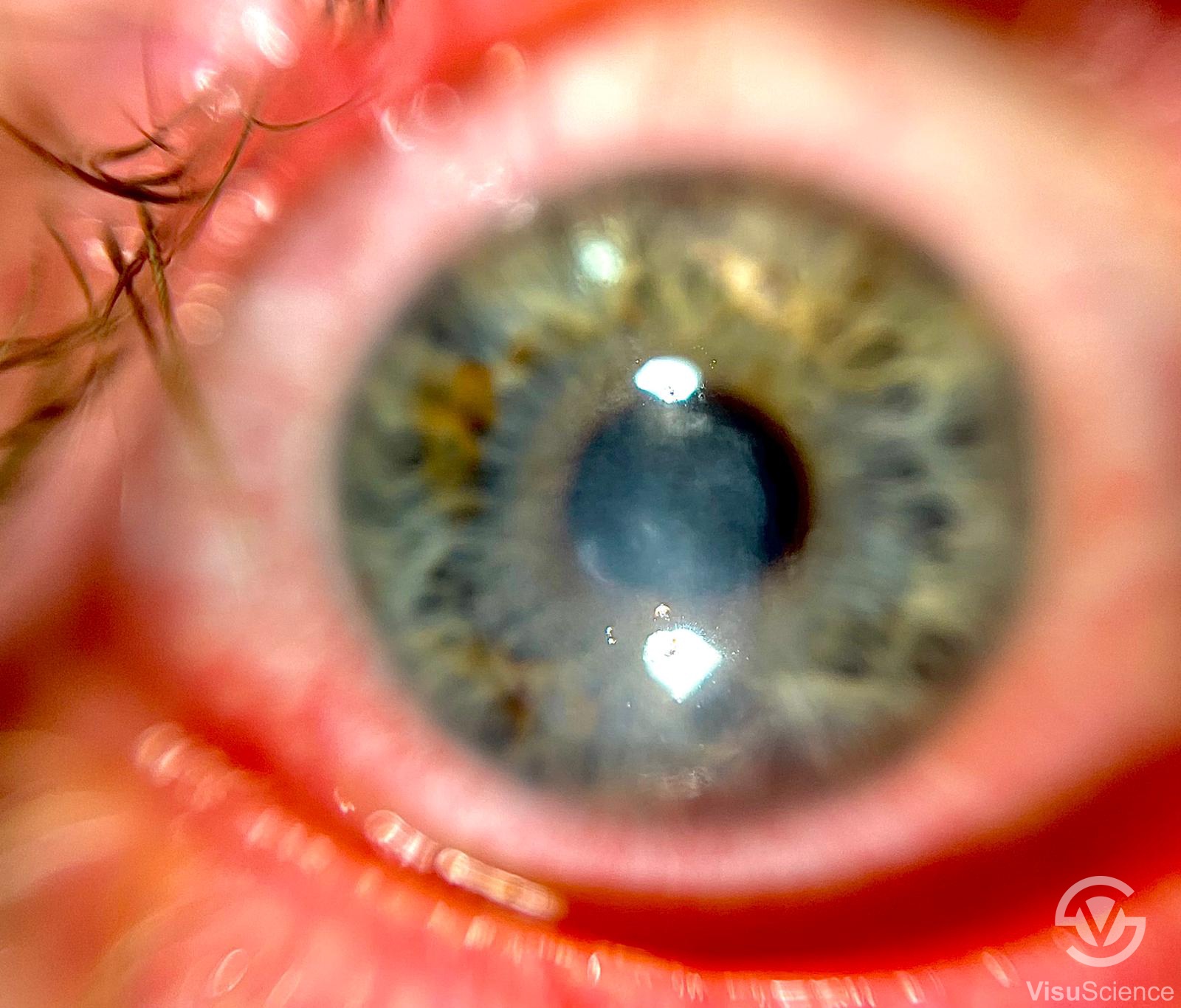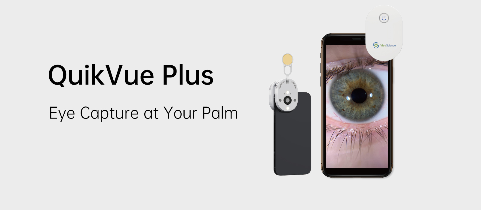
| QuikVue Plus Design Concept It is well acknowledged that imaging is playing a more and more important role in ophthalmology diagnosis. QuikVuePlus is a smartphone adaptor designed especially for anterior segment imaging. It provides 10x magnification which meets the basic eye examination demand for optometrists, general practitioners, pediatricians, veterinarians, etc. The specially designed optical lens enables QuikVue Plus to capture clear anterior images for primary eye care examination and telemedicine.
| ||||||||||||||||||||||||||||
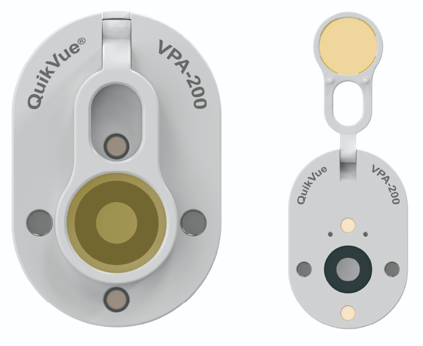
| White and Blue Illumination | ||||||||||||||||||||||||||||
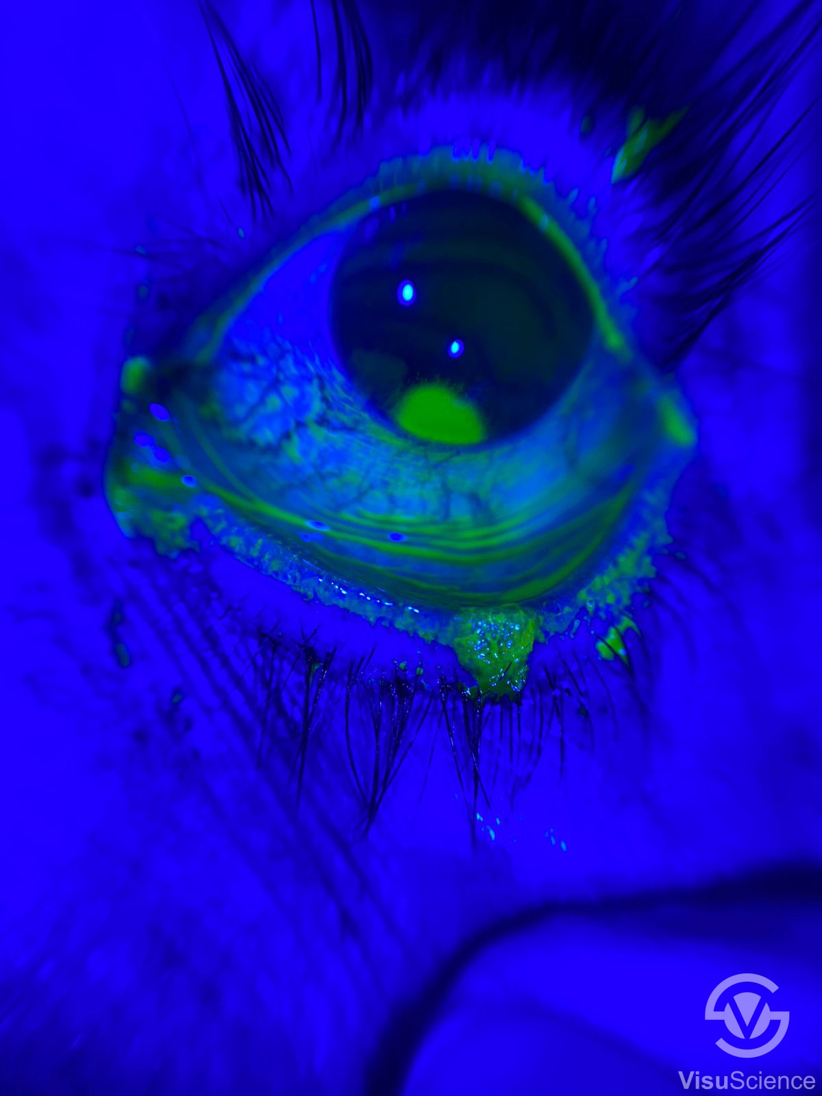
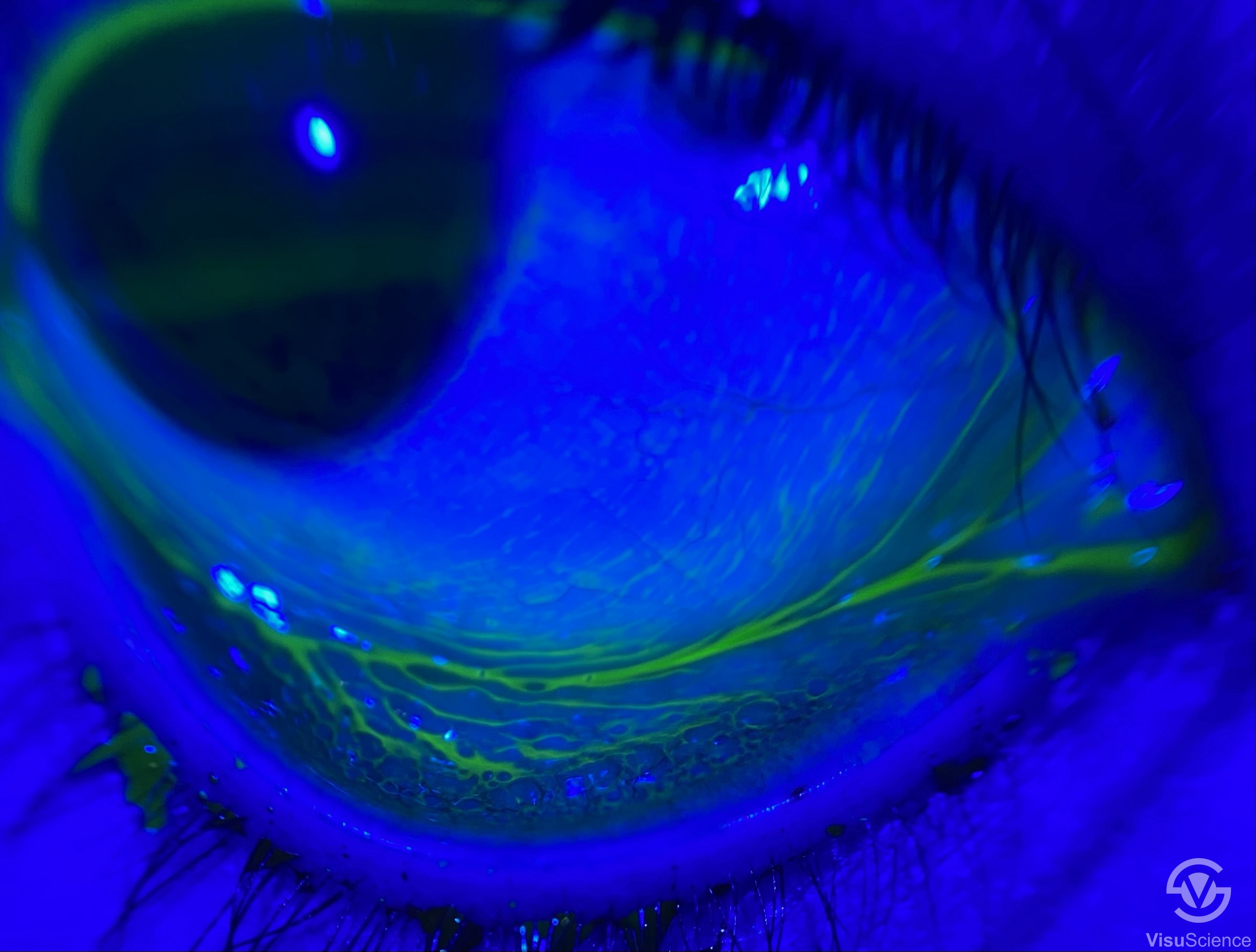
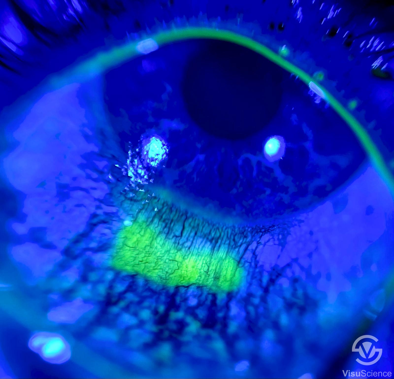
QuikVue Plus provides both white and blue illumination respectively. The white LED projects warm white light which is similar to slit lamp’s halogen illumination. There are two levels of white illumination available to meet different brightness demand during examination. The blue illumination can be used to capture fluorescein images to assist diagnosis with corneal staining and contact lens fitting, etc. | 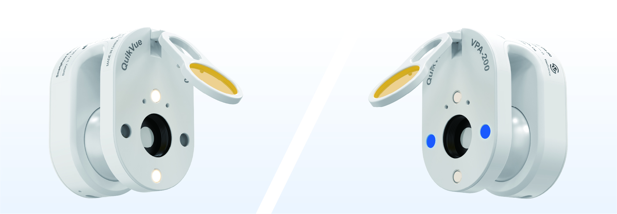 | |||||||||||||||||||||||||||
| Integrated Yellow Filter | ||||||||||||||||||||||||||||
| QuikVue Plus is equipped with an integrated yellow filter. The flipable yellow filter making it easy for clinicians to take enhanced fluorescein images with cobalt blue illumination. This function is especially useful in lens fitting, cornea ulcer imaging, tear film break up time examination, etc. | |||||||||||||||||||||||||||
| Smartphone Compatibility | ||||||||||||||||||||||||||||
Thanks to the air cushion design, QuikVue Plus is able to attach on most of the phones in the market firmly. Clinicians need to align QuikVue Plus lens with smartphone’s main camera. The requirements for the main camera location is as below: | ||||||||||||||||||||||||||||
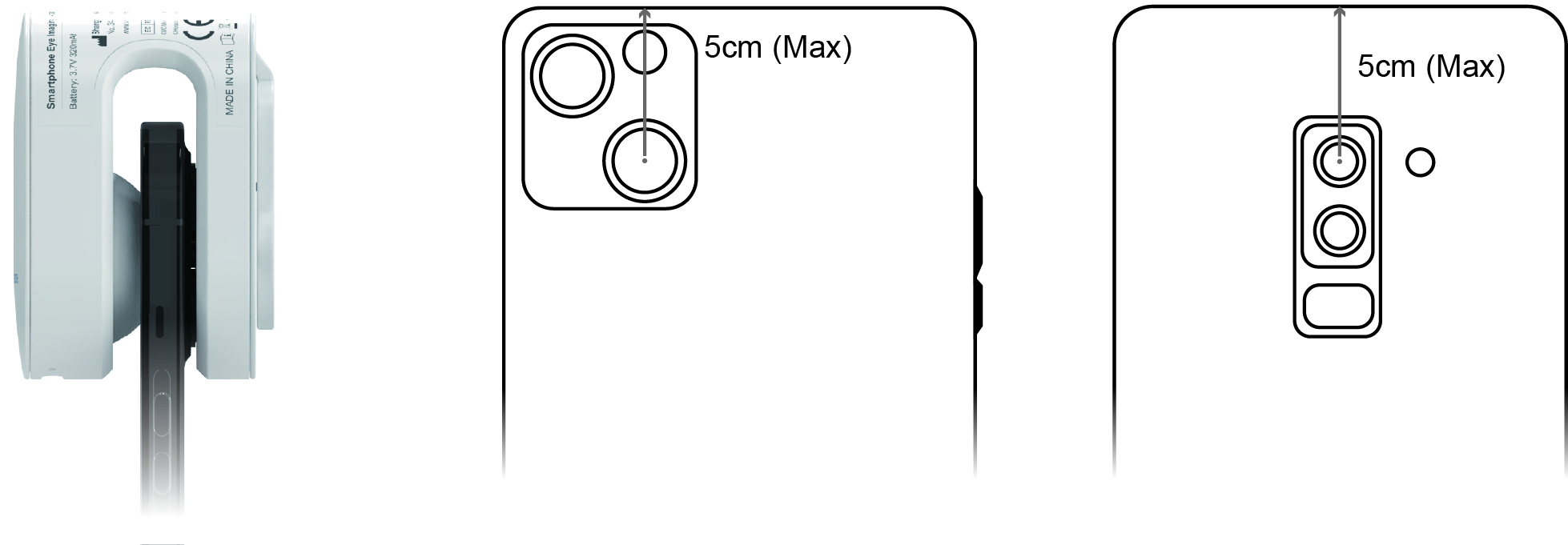 | |||
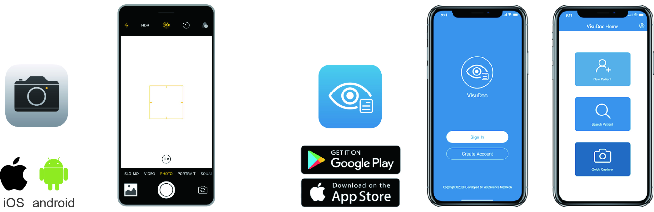
Working Application | |||
QuikVue plus can work with smartphone default camera app or VisuScience developed VisuDoc ophthalmic case management app. VisuDoc can be downloaded from both App store and Google play store. | |||
 | |||
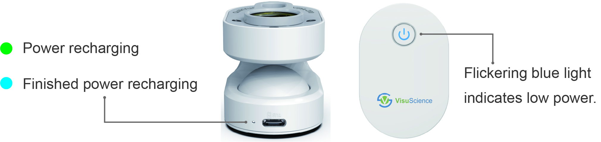
| Power Management | |||
QuikVue Plus applies mini Li-ion rechargeable battery. It can be recharged through a Type C cable. There is one light indicator next to the Type C charging port. When the power recharging is finished, the indicator light will turn from blue to green.
The switch button has blue light on when working normally. If the power is low, the light will flicker to remind clinicians to do recharge. | |||
 | |||
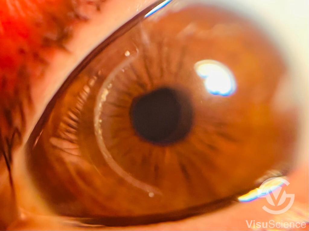
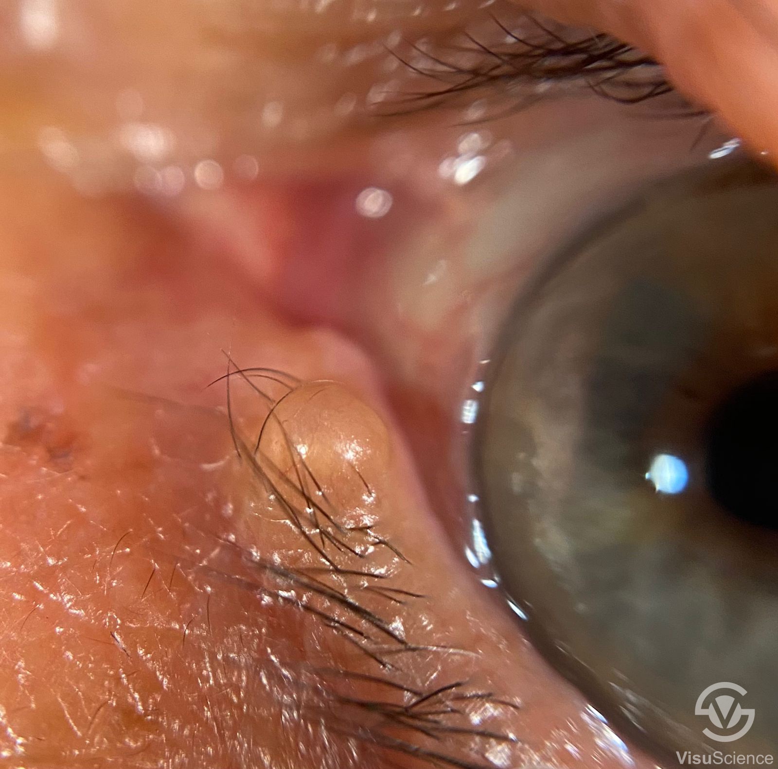
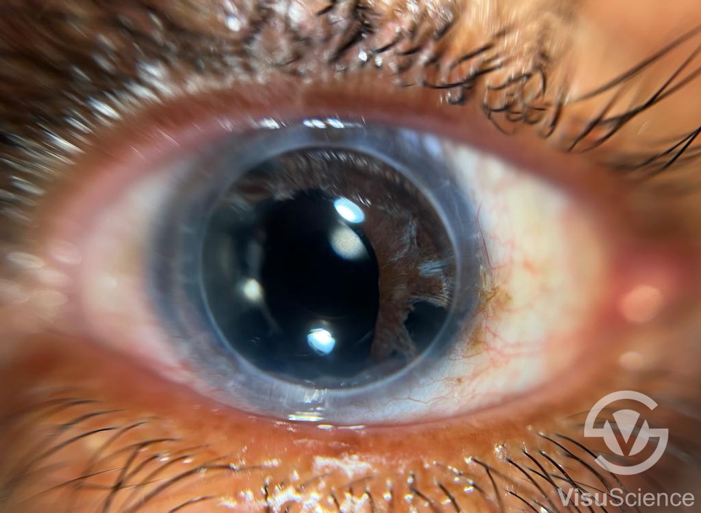
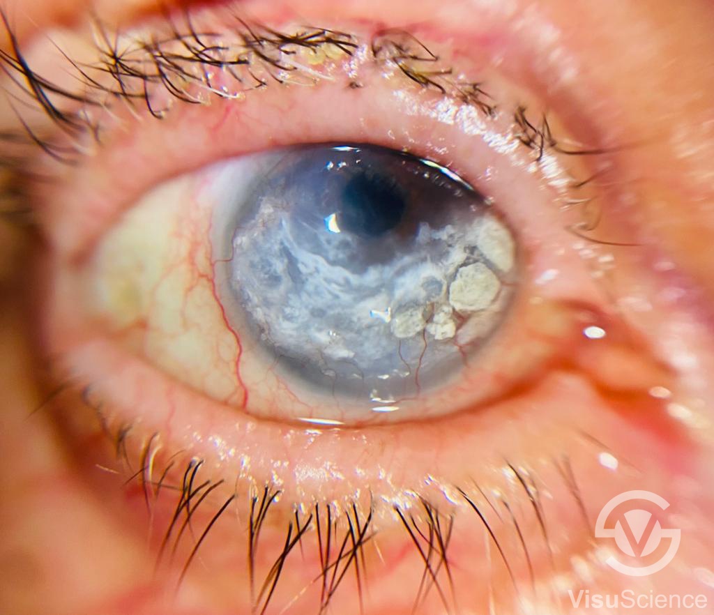
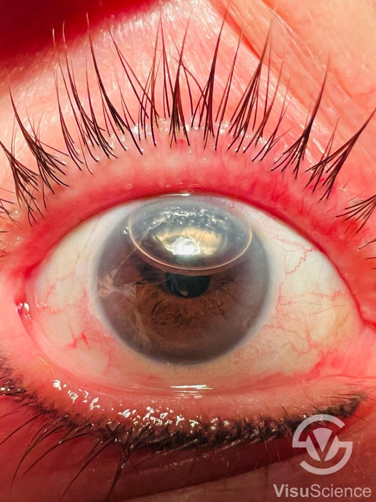
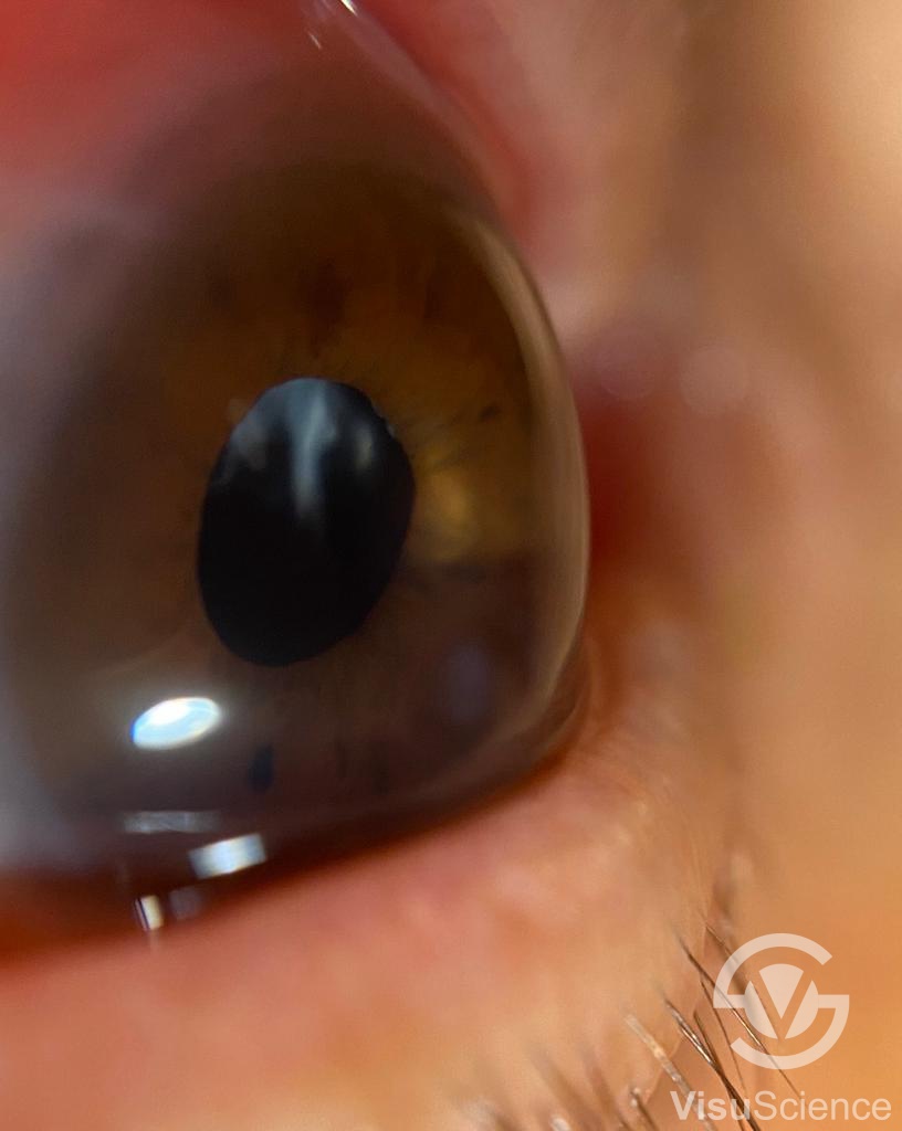
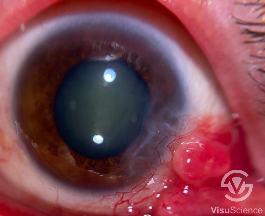
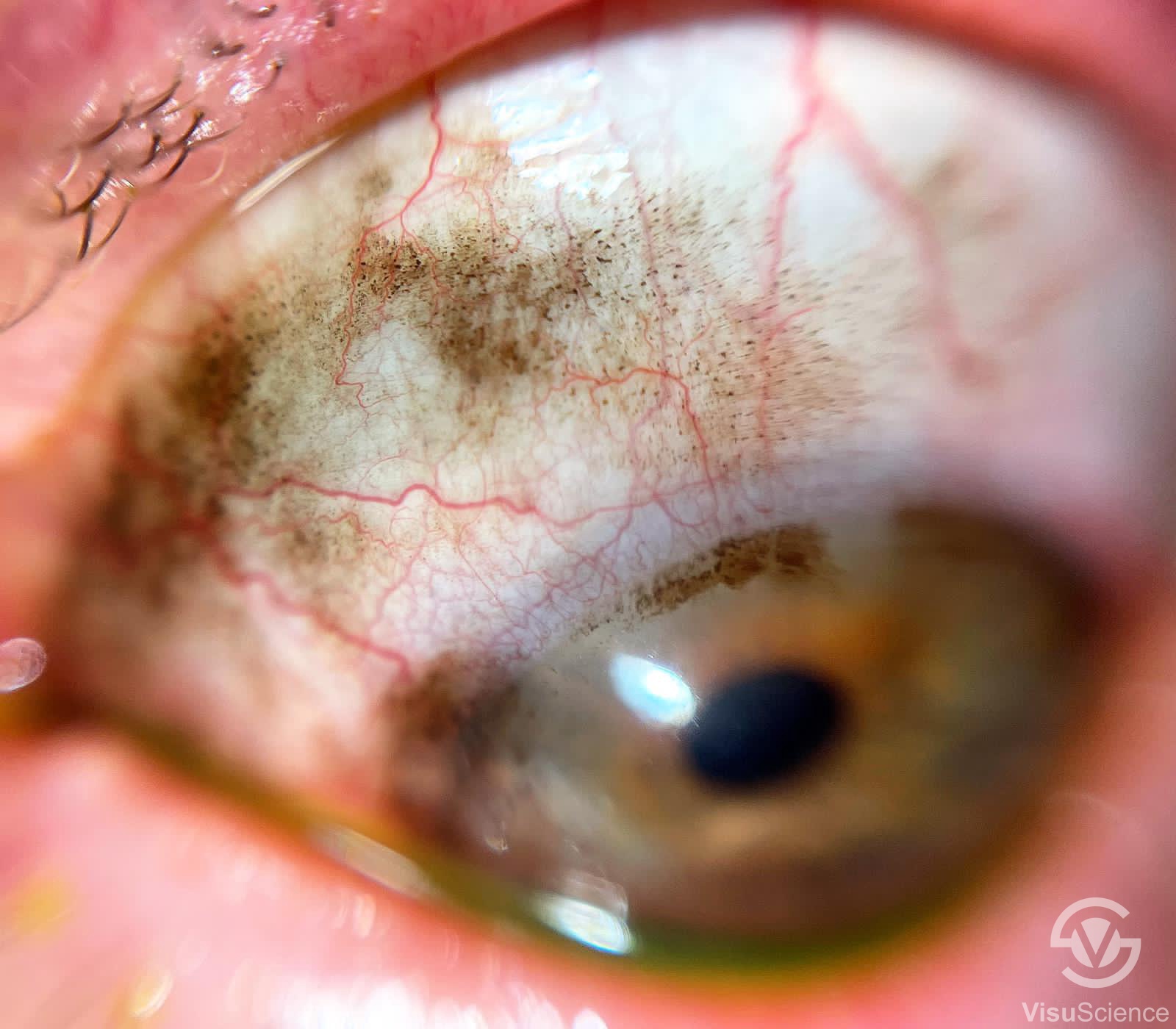
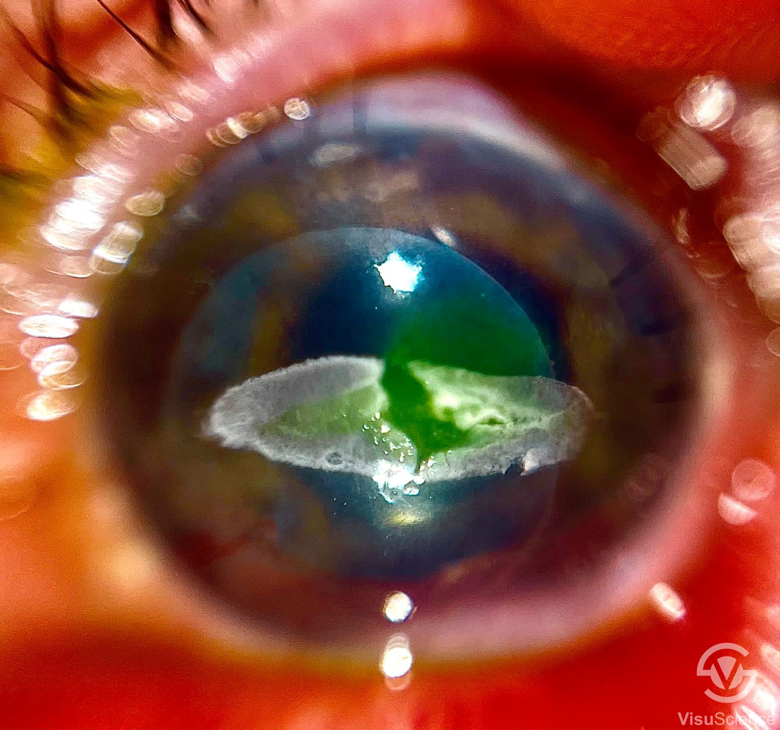
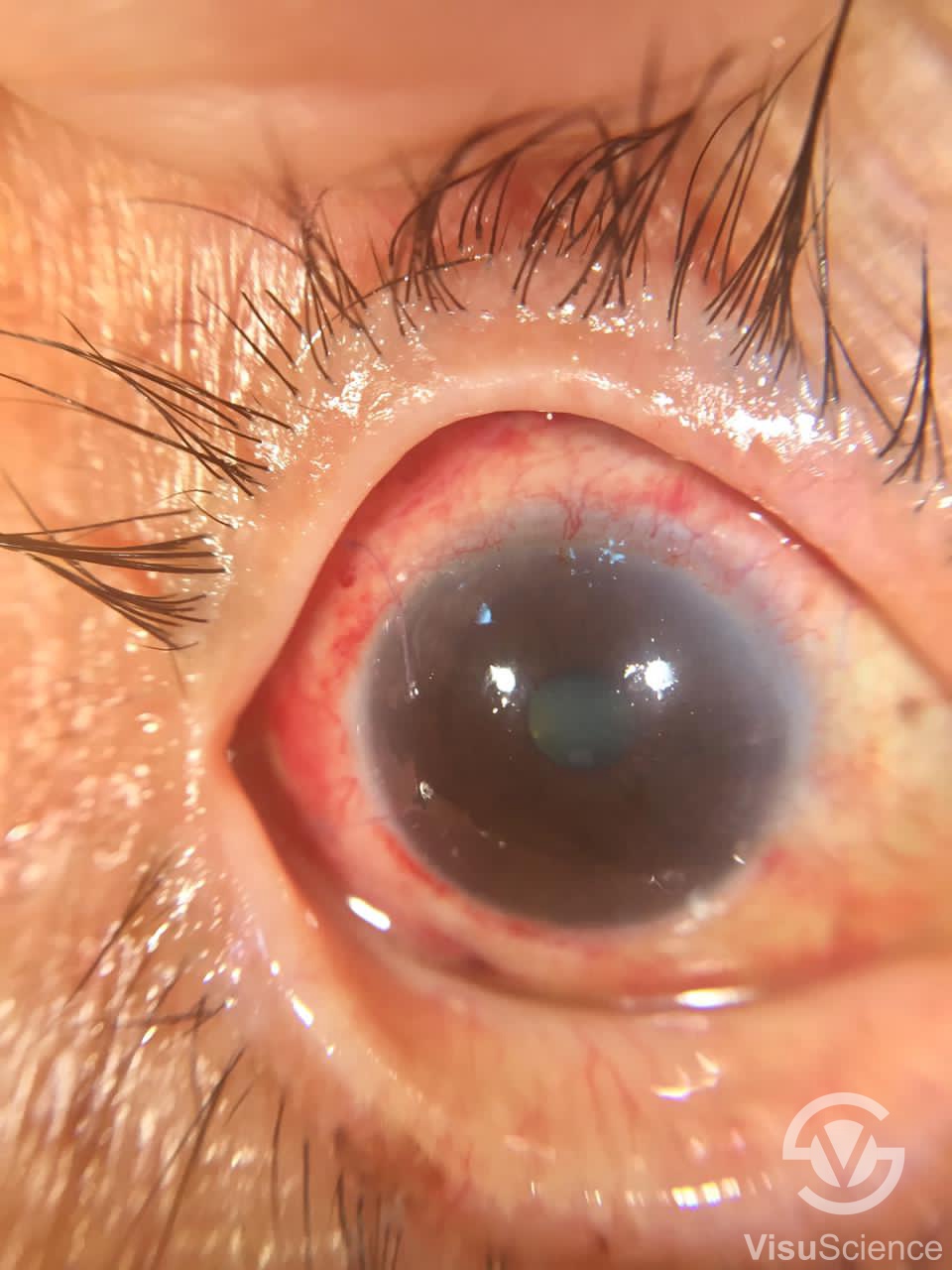
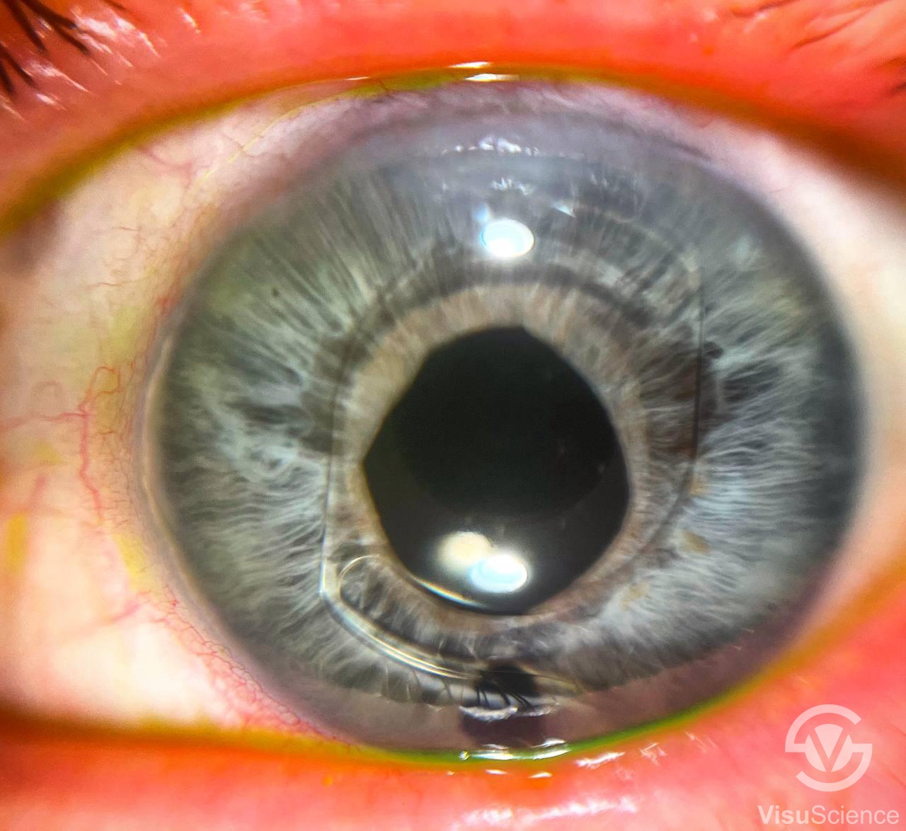
| Image Showcase | |||
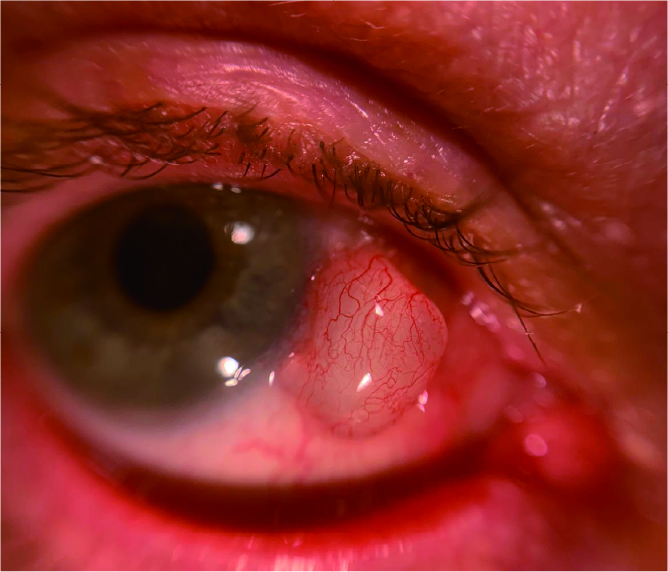 | 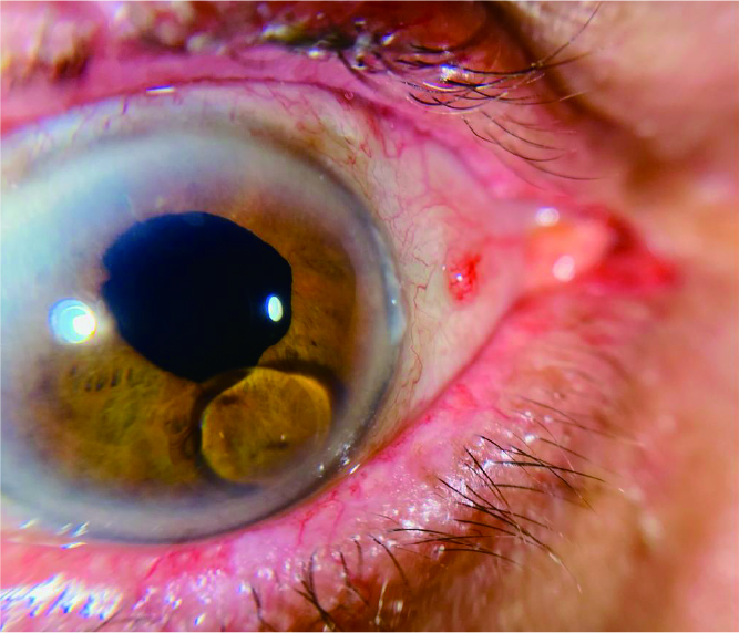 | 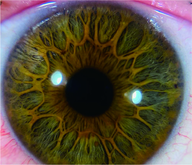 | 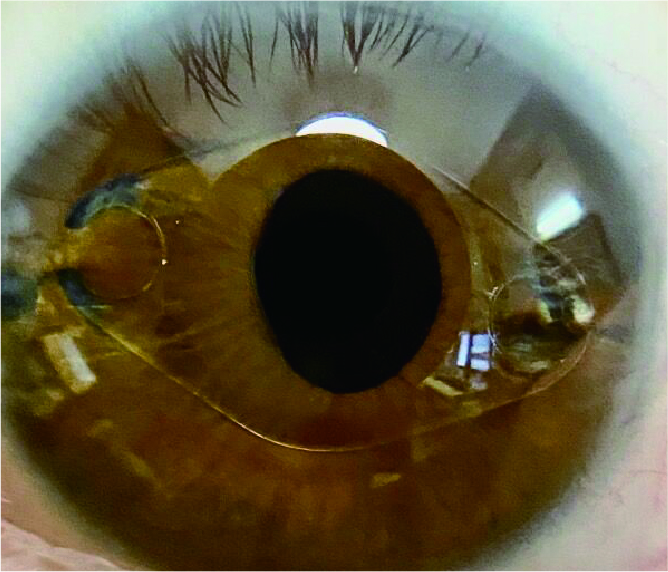 |
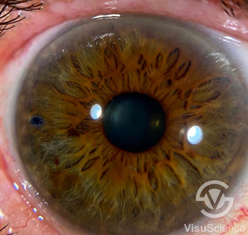
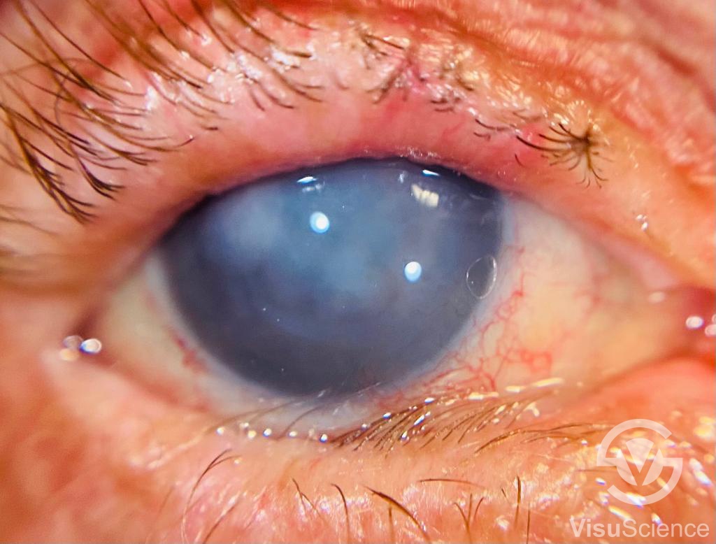
Corneal Cyst | silicone oil migration | Flower Iris Pattern | ICL Implant |
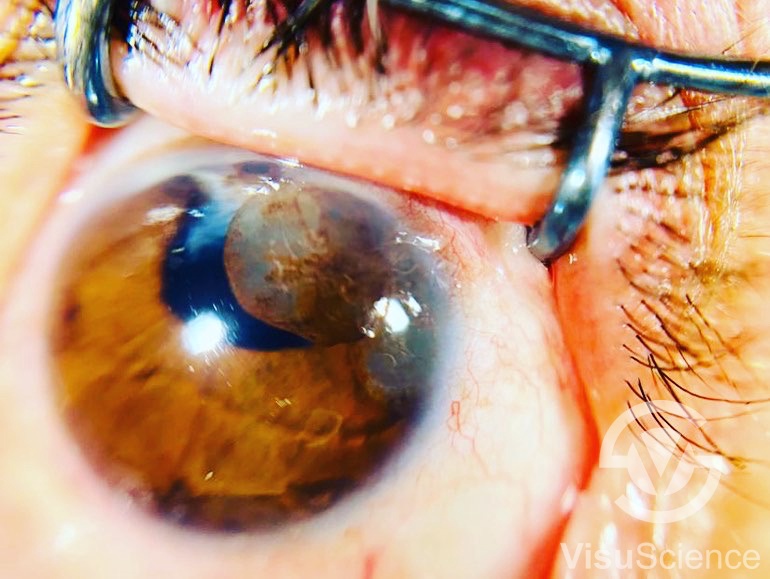
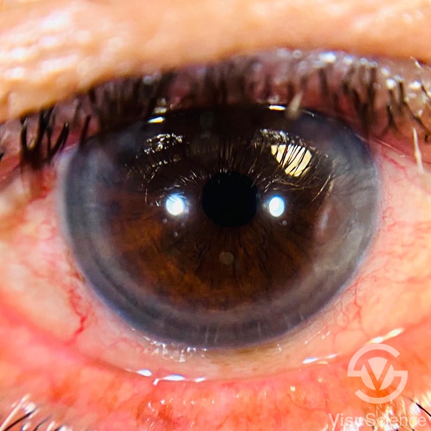
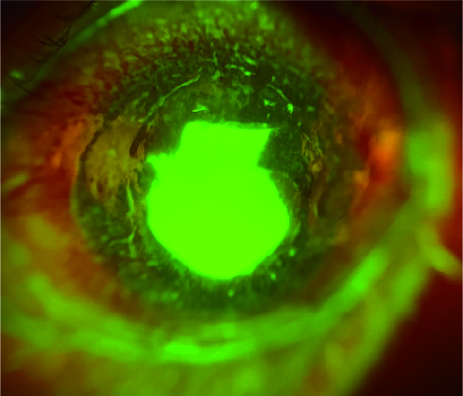 |  |  |  |
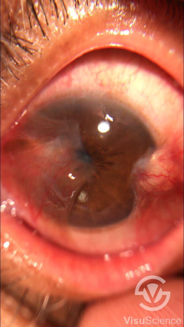
Corneal Ulcer | OK lens fitting | Marginal Keratitis | Dendritic ulcer |
Click here to see more images captured by QuikVueTM .
| Testimonial for QuikVueTM | |
| Dr. Hesham Mahmoud Eissa is a consultant ophthalmologist. He bought QuikVueTM at EOS (Egyptian Ophthalmic Congress) in March 2019. He is very happy with the device and here come his comments: | |
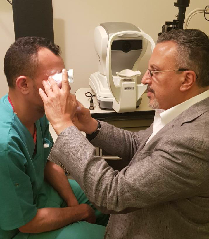 | “Your product is great and I have using it since with my patients... Needless to say, it is quite helpful in showing and explaining to patients in a far easier way the condition and showing also the improvement when it comes to corneal infections for example...” |
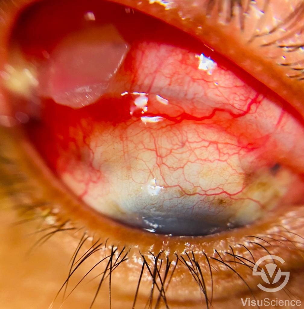
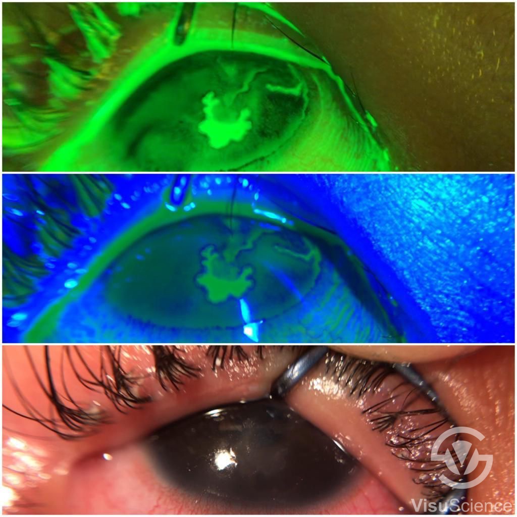
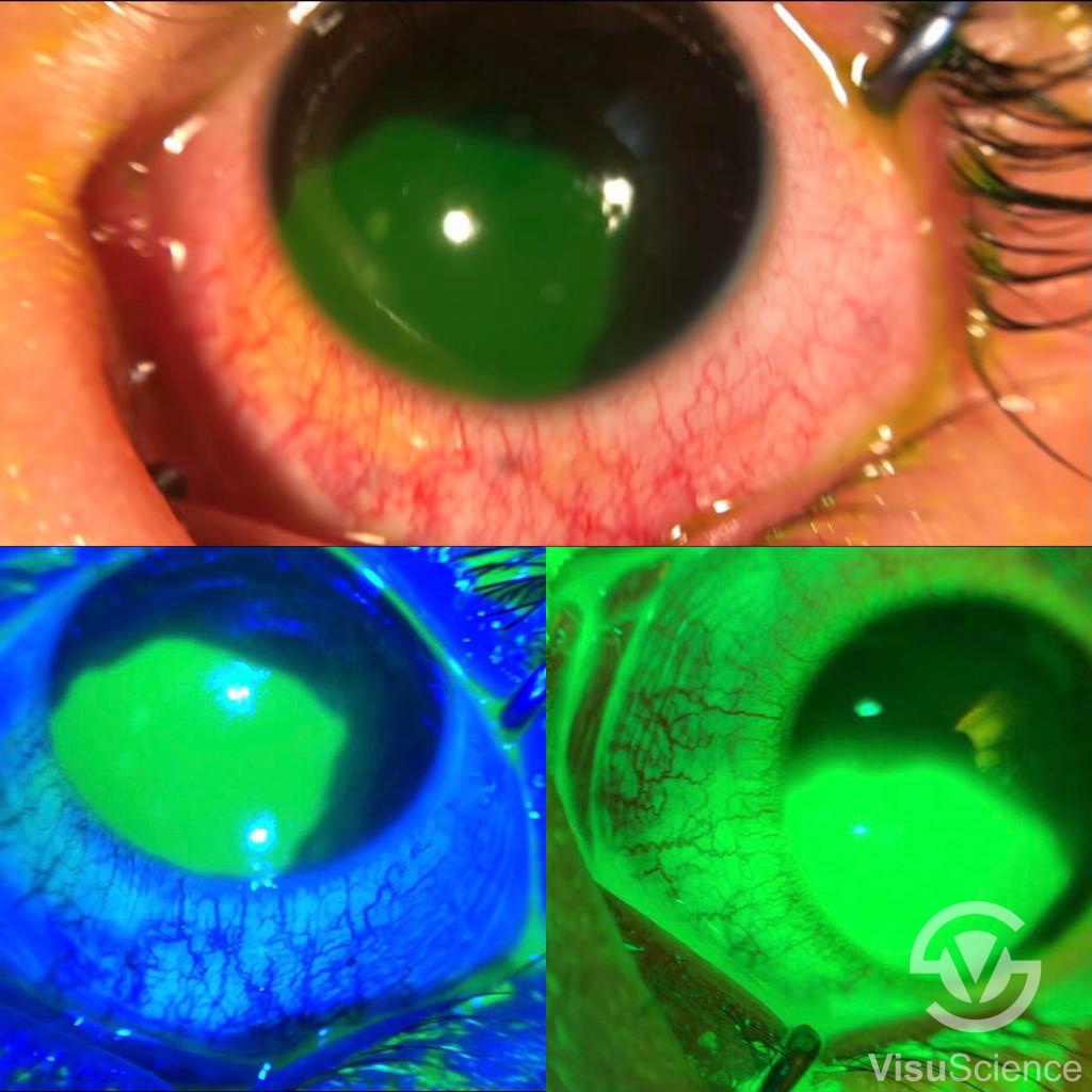
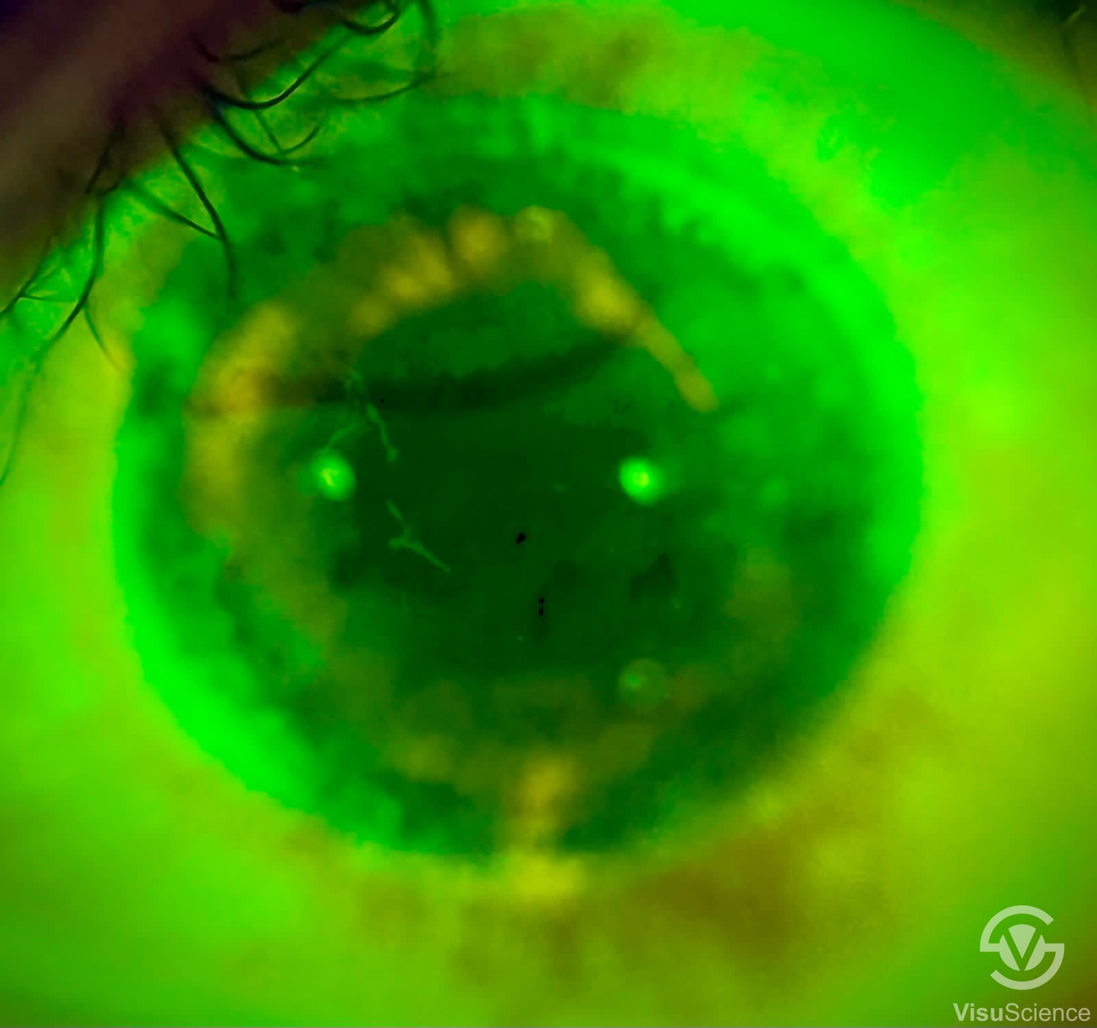
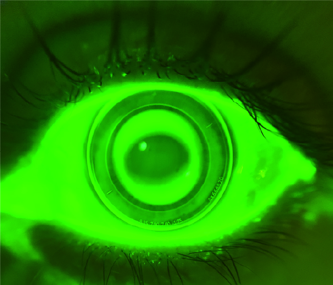
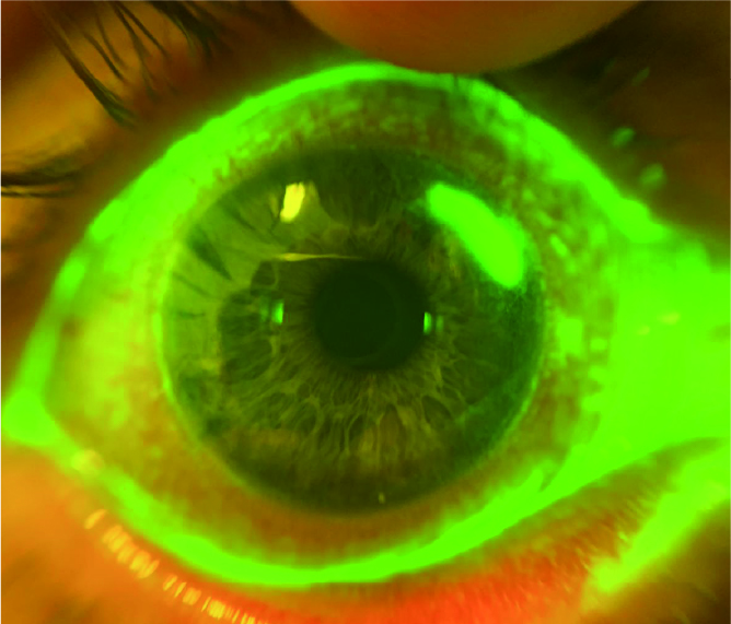
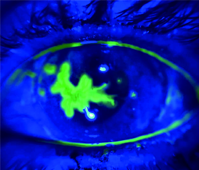
| Pathological Images Captured by Dr. Hesham Mahmoud Eissa |
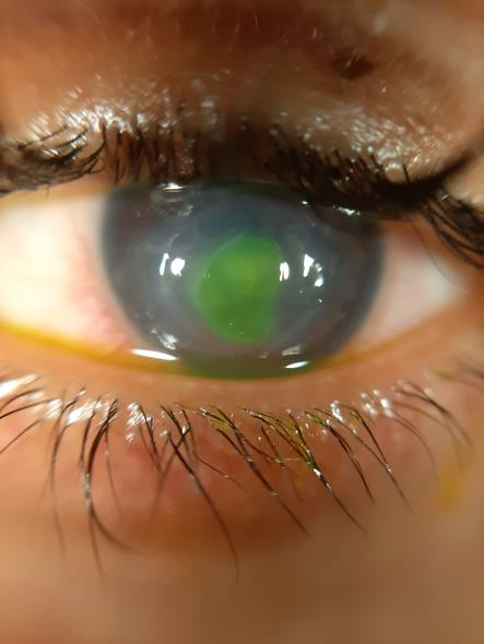 | 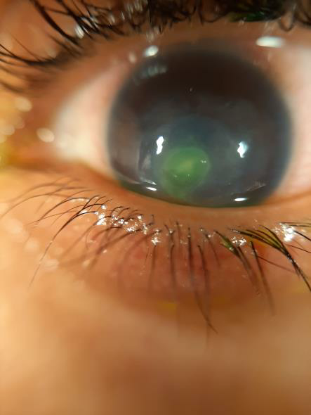 | 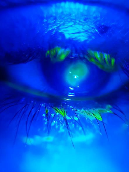 |
| Contact lens related corneal infection before empirical fortified eye drops treatment, notice the size of the ulcer stained with green | Same case after one week of treatment showing decrease in ulcer size | Same case after one week of treatment photo with blue filter |
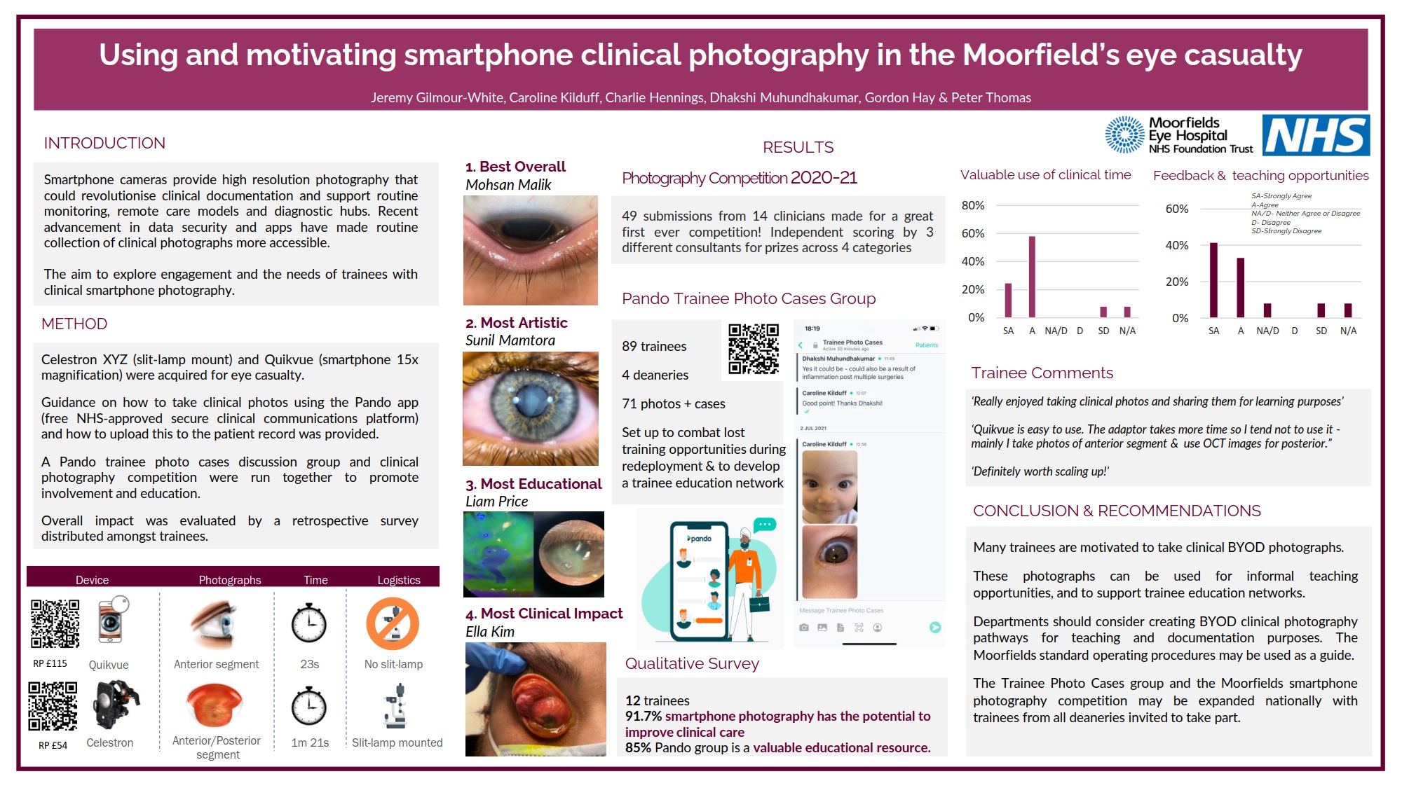
Clinical User Feedback from UK Moorfields Hospital Doctors
 | ||
| Related Products | |
 | |
Powered by Froala Editor